Home
Blood Typing
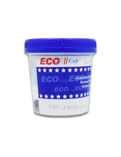
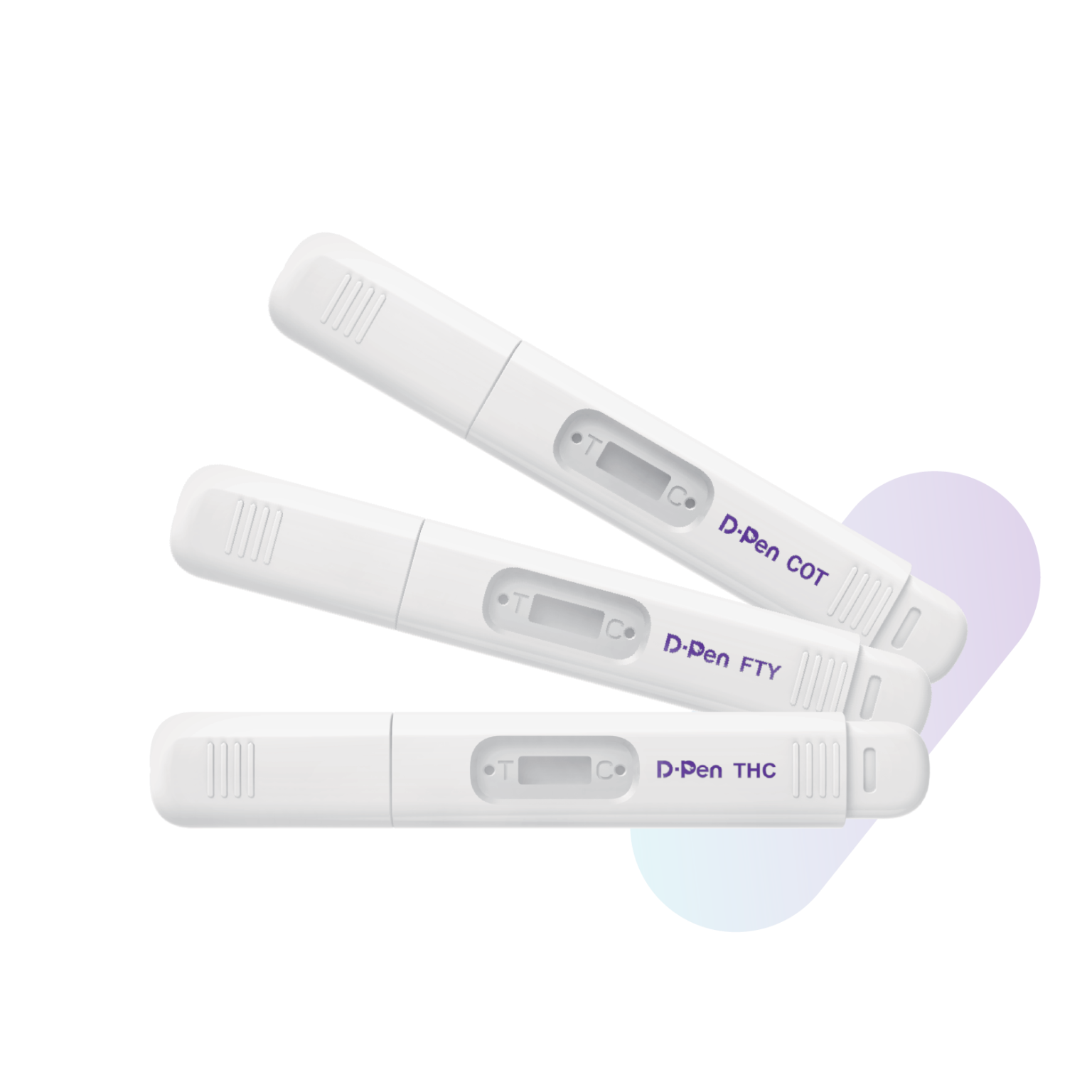
Drug Tests
Nicotine Tests
Alcohol Tests
Pregnancy
General Health
Cart
Home
Craig Medical Wholesale Distribution
Professional and at-home point-of-care diagnostic products
Professional Healthcare Industry • Government • Educational Institutions • Retailers • Work Place • Individuals
Featured Products
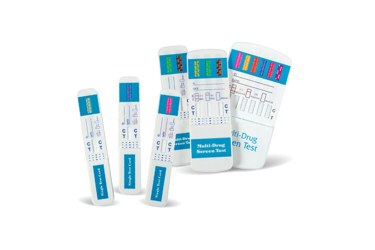
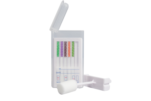
Drug Tests
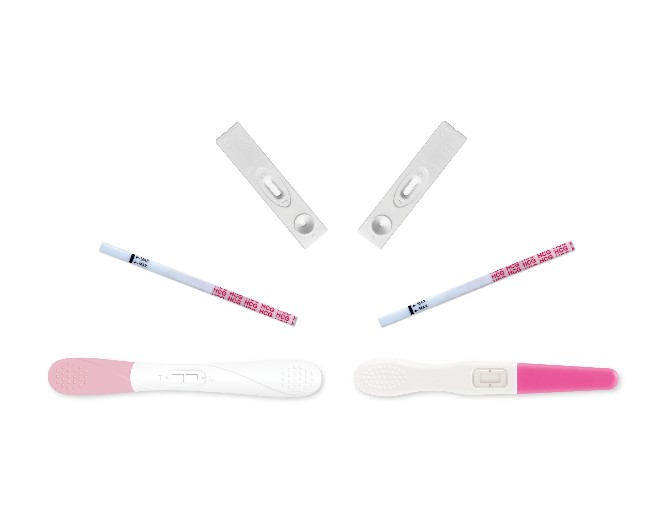
Pregnancy Tests
Categories
Testing Accessories
Drug Tests
Nicotine Tests
Alcohol Tests
Pregnancy & Fertility
General Health